
The short answer
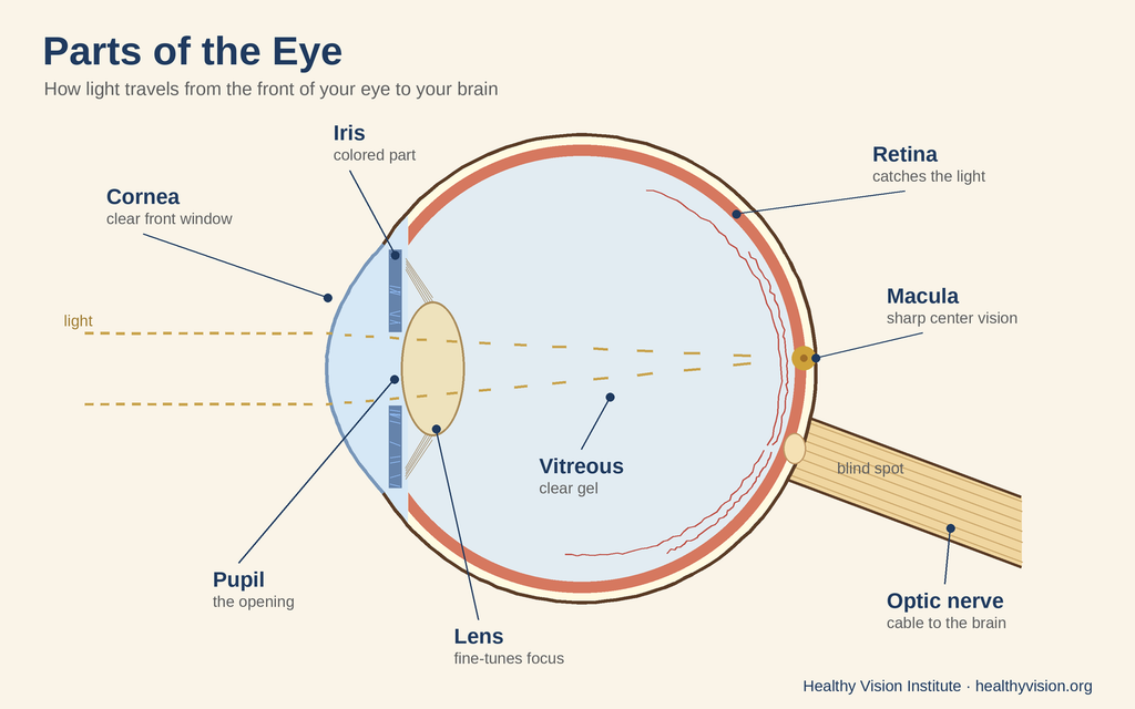
Your eye works like a camera. The cornea and lens in the front focus light. The iris and pupil control how much light gets in. The retina at the back catches the light and turns it into signals. The optic nerve carries those signals to your brain, and your brain turns them into the picture you see. Many eye problems start deep inside the eye with no pain, so an eye exam is the only way to know every part is healthy.
Right now, light from this screen is entering your eyes, getting focused, and turning into signals your brain reads as words. It happens in a split second, and you never think about it.
You don’t need a medical degree to understand how it works. Knowing the basic parts helps you follow what your eye doctor says at your exam. When you hear “your retina looks healthy,” you’ll know what that means — and why it matters.
Here is a simple tour of the eye, from front to back.
Key Takeaways
- Your eye works a lot like a camera: it focuses light and sends the picture to your brain.
- The cornea and lens focus light. The iris controls how much light gets in.
- The retina at the back catches the picture. The optic nerve carries it to your brain.
- Clear fluid and gel inside the eye let light pass through.
- Your brain does the last step and turns signals into the picture you see.
- An eye exam checks each part, often before you would notice a problem.
How Does the Eye Work? Think of a Camera
A camera has three main jobs: a lens to focus light, an opening that controls how much light comes in, and a sensor in the back that captures the picture. Your eye has all three.
Light comes in the front, gets focused, passes through the middle of the eye, and lands on the back wall. The back wall turns light into signals, and your brain turns those signals into the picture you see. The big difference from a camera: your eye adjusts itself all day, on its own.
The Front of the Eye
The cornea: the clear front window
The cornea is the clear, dome-shaped window at the front of your eye. It’s the first thing light hits, and it does most of the focusing. It has no blood vessels, which helps keep it clear. It is also full of nerve endings, which is why a tiny speck of dust feels so big. If the cornea’s shape is a little off, things look blurry — a common reason people need glasses or contacts.
The iris and pupil: the light controls
The iris is the colored part of your eye — blue, brown, green or hazel. It’s actually a muscle. The pupil is the black circle in the middle. It’s an opening that lets light in. In bright light, the iris makes the pupil smaller. In the dark, it makes the pupil bigger. A camera does the same thing to get the right amount of light.
The lens: the fine-tuner
Behind the iris is the lens. It’s clear and bendy, and it finishes the focusing the cornea started. Tiny muscles change its shape so you can switch from looking at a street sign to looking at your phone.
With age, the lens gets stiffer, so focusing up close gets harder. That’s why many people need reading glasses in their 40s. Over time, the lens can also turn cloudy. That’s called a cataract.
The Inside of the Eye: Clear Fluid and Gel
For light to reach the back of the eye, the inside has to be clear. Two clear fillings do this job.
Aqueous humor: the watery fluid in front
The aqueous humor (a clear, watery fluid) fills the space between the cornea and the lens. It feeds the front of the eye and keeps a steady pressure inside it. Your eye doctor checks this pressure at your exam, because pressure that’s too high is linked to glaucoma.
The vitreous: the clear gel that fills the eye
The vitreous is a clear gel that fills most of your eyeball and gives it its round shape. As you get older, the gel slowly shrinks and changes. That’s one reason people start to see floaters — little specks, dots or cobwebs that drift across your vision.
New floaters, flashes or a shadow? Call right away.
A few floaters that have been there a long time are common. But call your eye doctor right away — or go to the emergency room — if you suddenly see:
- A shower of new floaters
- Flashes of light
- A shadow or curtain moving across part of your vision
These can be signs of a torn or detached retina, which needs care fast.
The Back of the Eye
The retina: the eye’s camera sensor
The retina is a thin layer of tissue that lines the back of the eye. It’s covered in millions of tiny cells that catch light and turn it into electrical signals. There are two kinds. Rods help you see in dim light and at the sides. Cones help you see color and fine detail.
Many serious eye problems happen in the retina. For example, diabetes can damage the tiny blood vessels there. This is called diabetic retinopathy, and it often has no signs early on. That’s why people with diabetes need a dilated eye exam (an exam with drops that widen your pupils) at least once a year.
The macula: your sharp center vision
The macula is a small spot in the center of the retina. It gives you the sharp, straight-ahead vision you use to read, drive and see faces. The very center of the macula, called the fovea, gives you your clearest vision of all. The rest of the retina handles your side vision. When the macula breaks down, it’s called macular degeneration.
The optic nerve: the cable to your brain
The optic nerve carries signals from the retina to your brain, like a cable from a camera to a computer. Everything you see travels down this nerve. Glaucoma damages the optic nerve, often slowly and without pain.
Your blind spot
Where the optic nerve leaves the eye, there are no light-catching cells. That makes a small blind spot in each eye. You never notice it, because your brain fills in the gap using what your other eye sees.
The Muscles That Move Your Eyes
Six small muscles on the outside of each eye move it up, down, side to side and at an angle. Both eyes have to point at the same thing at the same time. That teamwork lets your brain mix two slightly different pictures into one picture with depth. When the muscles don’t work together well, it can cause eye strain, double vision, or an eye that turns in or out.
Tears, Eyelids and Lashes: The Eye’s Protection
Your eyelids work like windshield wipers. Every blink spreads a fresh layer of tears across your eye and sweeps away dust. Your lashes and brows help keep out dirt and sweat.
The thin layer of tears on your eye is called the tear film. It has a watery layer and a thin layer of oil on top that keeps the tears from drying up too fast. When the tear film doesn’t work well, your eyes can feel gritty, burning or tired. That’s dry eye.
How It All Works Together
Say you look up at a bird in a tree. Here’s what happens:
- Light bounces off the bird and goes through your cornea, which starts to focus it.
- Your iris makes the pupil the right size for the light.
- Your lens changes shape to make the bird sharp.
- The light passes through the clear vitreous gel.
- It lands on your retina. Your macula picks up the fine detail.
- Your optic nerve carries the signals to your brain.
- Your brain turns the signals into the picture of the bird you see.
All of that happens in a split second, over and over, all day long. Every part has to do its job.
Why This Matters for Your Eye Health
Your vision depends on every part of the eye being healthy. A problem in any one part can change what you see. That’s why a full eye exam checks the front of the eye, the pressure inside, the lens, the retina and the optic nerve.
Here’s the most important part: many eye problems start with no pain and no signs, deep inside the eye where you can’t feel them. Glaucoma, early macular degeneration and diabetic eye disease often work this way. An eye exam is the only way to know for sure that every part is healthy — even if your vision feels fine.
Due for an eye exam? We see patients in St. Petersburg, Clearwater and New Port Richey, and we check your insurance before your visit.
Frequently Asked Questions
What are the parts of the eye and their functions?
The main parts are the cornea (clear front window), iris (colored part), pupil (the opening in the middle), lens (the fine-tuner), vitreous (the clear gel inside), retina (the light-catching layer at the back), macula (the center of the retina), and optic nerve (the cable to the brain).
What part of the eye actually sees?
No one part does it alone. The retina turns light into signals, the optic nerve carries them, and your brain builds the picture. Seeing is teamwork between your eyes and your brain.
What does the cornea do?
The cornea is the clear front of your eye. It lets light in and does most of the focusing. If its shape is a little off, things look blurry, and glasses or contacts can help.
What’s the difference between the cornea and the lens?
Both focus light. The cornea sits at the very front and does most of the focusing. The lens sits behind the iris and changes shape to fine-tune, so you can switch between near and far.
What is the macula?
The macula is a small spot in the center of the retina. It gives you sharp center vision for reading, driving and seeing faces.
What is the vitreous?
The vitreous is the clear gel that fills most of the eye and keeps its round shape. It changes as you age, which can cause floaters.
What causes floaters?
Most floaters are tiny clumps in the vitreous gel that cast shadows on the retina. They get more common with age. A sudden shower of new floaters, flashes of light, or a shadow across your vision needs care right away.
Why do my pupils change size?
Your iris makes the pupil smaller in bright light and bigger in dim light. It controls how much light gets in, and it happens on its own.
Why is it harder to see up close as I get older?
The lens gets stiffer with age, so it can’t change shape as easily to focus up close. That’s why many people need reading glasses in their 40s. It’s a normal part of aging.
Why do I need glasses if my eyes are healthy?
Glasses fix how light is focused. If the shape of your eye, cornea or lens makes light land a little in front of or behind the retina, things look blurry. Glasses bend the light so it lands in the right spot. Needing them doesn’t mean your eyes are unhealthy.
Can eye problems happen without any symptoms?
Yes. Glaucoma, early macular degeneration and diabetic eye disease can start with no signs. An eye exam can find changes before you notice them.
How often should I get my eyes checked?
It depends on your age, health and risk. People with diabetes need a dilated eye exam at least once a year. Your eye doctor will tell you how often is right for you.
See the Bigger Picture — Schedule an Eye Exam
Now you know how your eyes work. The next step is making sure every part stays healthy. A full eye exam checks your cornea, lens, eye pressure, retina and optic nerve.
At Healthy Vision Institute, Dr. James Powers and our team care for eyes in St. Petersburg, Clearwater and New Port Richey. Whether you’re overdue for a checkup or just want peace of mind, we’re here to help.
Ready to schedule your eye exam?
Sources: National Eye Institute (nei.nih.gov) — How the Eyes Work; Floaters; Retinal Detachment; Diabetic Retinopathy. American Academy of Ophthalmology (aao.org) — Parts of the Eye.
This article is for general education and is not medical advice. Talk with your eye doctor about your own eyes.